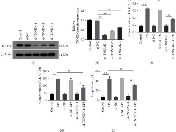
Figure 3.

Silencing TNNI3K alleviated LPS-induced H9C2 cell injury. (a) After knockout TNNI3K, the mRNA expression of TNNI3K in H9C2 cells was determined by qRT-PCR. (b) After knockout TNNI3K, the protein expression of TNNI3K in H9C2 cells was determined by western blot. (c) The activity of creatine kinase in different was detected by ELISA. (d) The LDH release activity in different was detected by LDH assay. (e) The mean fluorescence intensity of TUNEL staining in different groups. ∗ indicates comparison with the control group; # indicates comparison with the LPS group; & indicates comparison with the si-TNNI3K-1 group.
