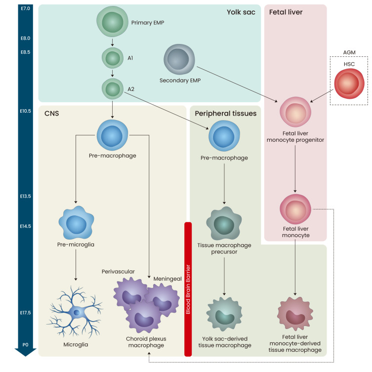
Fig. 1. Embryonic development of brain-resident macrophages.
Between embryonic day 7.0 (E7.0) and E8.0 of mouse development, primary erythromyeloid progenitor (EMP) cells in the yolk sac (YS) generate YS macrophages (A1 and A2), which are able to produce all types of tissue-resident macrophages including the brain. Around E10.5, YS macrophages move to central nervous system (CNS) or peripheral regions, and can differentiate into microglia or non-parenchymal macrophages (perivascular, meningeal, or choroid plexus macrophages) in the CNS or YS-derived tissue macrophages in the peripheral tissues. Secondary EMP cells in the YS and hematopoietic stem cells (HSCs) in the aorta-gonad-mesonephros (AGM) of embryo migrate to fetal liver during E8.5-10. Then, this fetal liver progenitor cells differentiate into fetal liver monocytes, which then invade all peripheral tissues except CNS at E14.5 of embryonic development.

