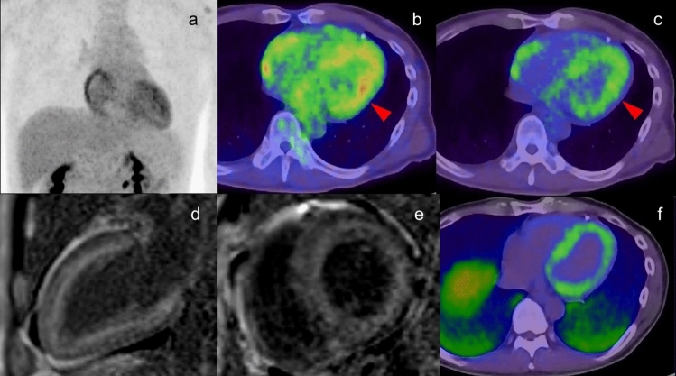
Fig.15.
Cardiac amyloidosis. Moderate FDG uptake was confirmed in left ventricle wall in the patient with AL amyloidosis (a, b red arrowhead). Different color scale of FDG PET/CT imaging demonstrated that FDG uptake existed in inner layer of left ventricle wall, which were matched to the area with high intensity in PSIR MRI image (d, e) and the uptake in PiB PET/CT imaging (f) suggesting the amyloid deposit. The FDG uptake in the right atrium was cause by overload pressure of right atrium

