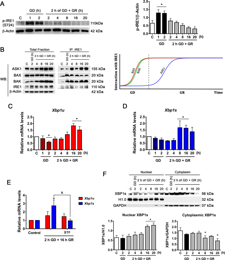
Fig. 2. IRE1α signaling activation during GD and GR.
A Representative immunoblot and quantification of p-IRE1α/β-Actin. B Analysis of the interaction of BAK, BAX, and ASK1 with IRE1α by co-immunoprecipitation and detected by immunoblot. Representative immunoblots from two independent experiments and a diagram of the interaction kinetics of BAX, BAK, and ASK1 with IRE1α along the GD and GR periods. Expression levels of C Xbp1u and D Xbp1s mRNA levels as determined by qRT-PCR in cortical neurons exposed to GD/GR. E Effect of the inhibition of IRE1α RNAse activity on Xbp1u and Xbp1s expression. IRE1α RNase activity inhibitor (STF, 30 µM) was incubated during GR. F Analysis of the subcellular localization of processed XBP1 (XBP1s). Representative immunoblot and quantification of XBP1s in the nucleus and cytoplasm. Data represent the mean ± SEM of 5 (A), 4 (C, D), and 3 (E, F) independent experiments and were analyzed by one way ANOVA followed by Fisher’s multiple comparisons, *p < 0.05 vs. control, &p < 0.05 vs. 2 h GD + 16 h GR.

