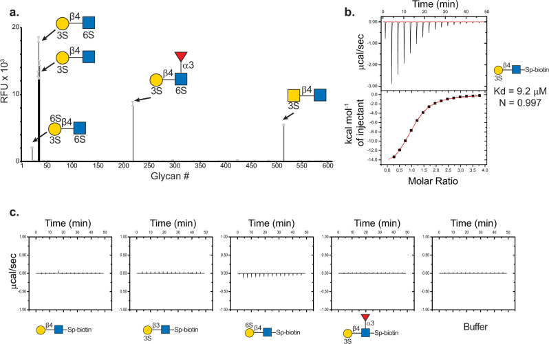
Fig. 1. The specificity of VLR O6 as determined by glycan microarray and ITC.
a O6-mFc chimeric protein screened on CFG mammalian glycan microarray at 10 µg/mL reveals the binding motif of O6 is (3 S)Galβ1-4GlcNAc. RFU relative fluorescence units, error bars = ±1 SD. b Monomeric VLR O6 binds to ligand with ~10 µM affinity. c No detectable binding curves were observed by ITC for the following structures: Galβ1-4GlcNAc, (3 S)Galβ1-3GlcNAc, (6 S)Galβ1-4GlcNAc and (3 S)Galβ1-4(Fucα1-3)GlcNAcβ-Sp. Yellow circle = Gal, yellow square = GalNAc, blue square = GlcNAc, red triangle = fucose, 3 S and 6 S = position of sulfate.

