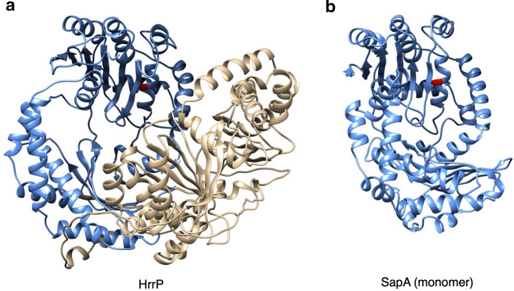
Figure 2.
Predicted structures of HrrP and SapA. (a) Structures of HrrP and SapA were modelled with I-Tasser and rendered by Chimera (closest PDB structural analogs for each protein were ID codes 6OFS and 1HR6 respectively). The two roughly symmetrical domains in HrrP are colored in blue and gold. (b) Though SapA is predicted to function as a homodimer it is depicted above in its monomeric form, colored blue. The active sites in both HrrP and SapA (E62 and E50, respectively), are shown in red.

