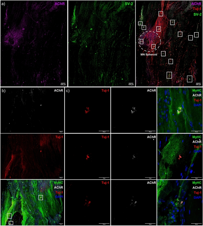
Figure 6.
Co-localisation of pre- and post-synaptic membrane proteins, motor nerve axons and myosin heavy chain positive fibres indicate neuromuscular junction formation in engineered tissues. (a) Confocal tile scan of neuromuscular tissue detailing AChR (left) and SV-2 (middle), with overlay (right) evidencing multiple synaptic contacts (outlined—white boxes) via co-localisation of pre- and post-synaptic markers and Tuj-1 + neuronal axons. (b) Confocal tile scan of neuromuscular tissues evidencing AChR, Tuj-1 + nerve terminal and myosin heavy chain co-localisation. (c) Zoom of synaptic contacts highlighted in (b) via white box outline. Images were collected across n = 8 neuromuscular tissues. Scale bars = (a, b) 100 µm, (c) 50 µm.

