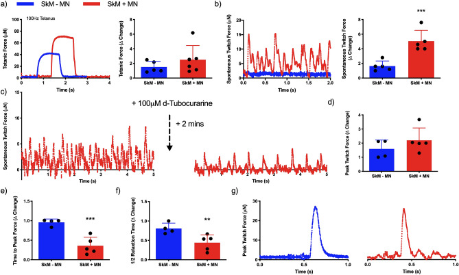
Figure 7.
Physiological functionality of engineered tissues is enhanced via neuromuscular junction formation. (a) Representative maximal induced tetanus contraction profiles of muscle (SkM -MN) and neuromuscular tissue (SkM + MN) and quantification of tetanic force. (b) Spontaneously occurring twitch force profiles in engineered tissues with (SkM + MN) and without (SkM–MN) iPSC motor neuron spheroids, and quantification of spontaneous twitch force driven via motor nerve inclusion. (c) Inhibition of the acetylcholine receptor in spontaneously contracting neuromuscular tissues via addition of 100 µM d-Tubocurarine. (d) Peak twitch force induced via electrical field stimulation. (e) Time to peak twitch force and (f) half relaxation time as physiological measures of contractile function. (g) Representative twitch profiles evidencing reduced time to peak twitch and shorter relaxation profiles in SkM + MN tissues. Δ change indicative of normalised data to SkM–MN. Beginning of representative tetanus and twitch contractions represents initiation of electrical field stimulation, time elapsed before this point is un-stimulated baseline recording. Individual functional data points indicative of n = 3 contraction profiles per tissue, totalling minimum of n = 12 and maximum of n = 18 contractions per condition and presented ± standard deviation (SD). **P ≤ 0.01 and ***P ≤ 0.001.

