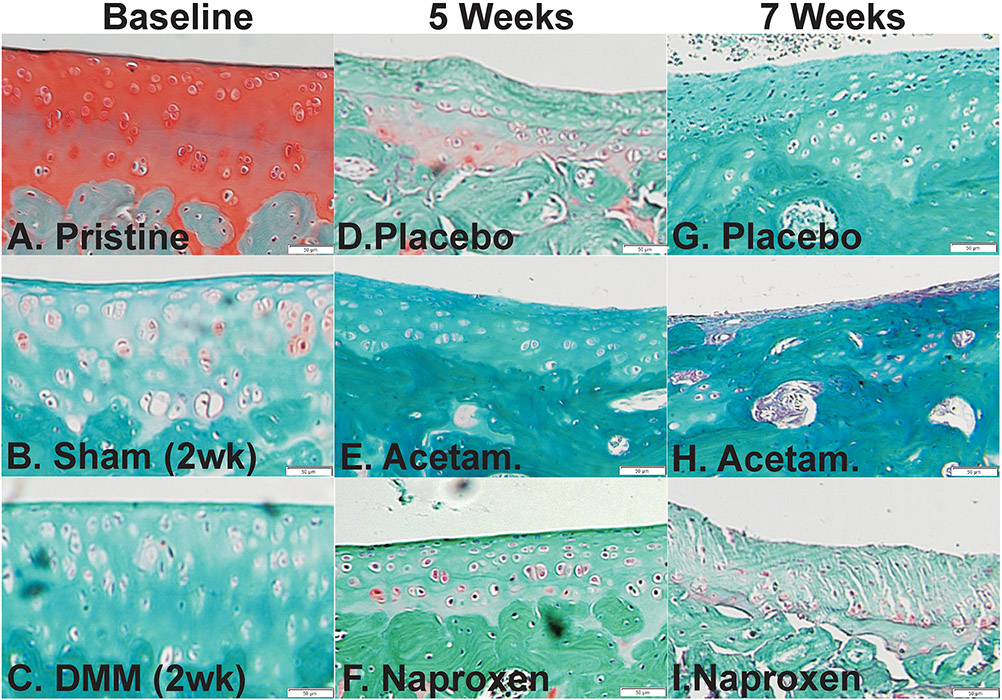
Figure 1.
Histological changes in the medial tibia cartilage following surgery and drug treatment. Shown are histology sections stained with safranin-O and fast green from pristine (A), baseline (2 weeks after surgery) sham (B), and baseline DMM (C) rat specimens. Histological sections from placebo (D, G), acetaminophen (E, H), and naproxen (F, I) treated rats are shown from specimens collected at 5 (D-F) and 7 weeks (G-I) after DMM surgery. Scale bar is 50 μm.

