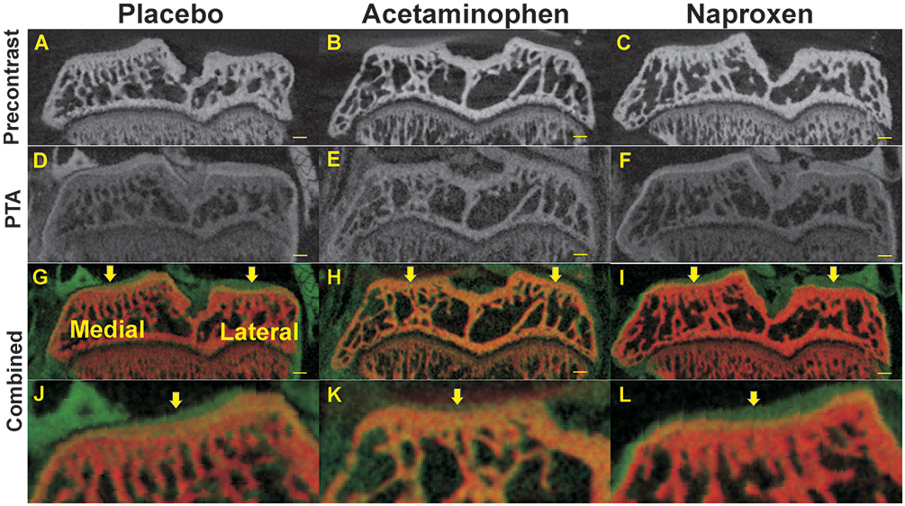
Figure 3.

Radiographic changes in tibia plateau cartilage and subchondral bone at 7 weeks after DMM surgery. Shown are pre-contrast (A-C), PTA post-contrast (D-F), and co-registered μCT images (combined, G-I) from rats treated with placebo (A, D, G, J), acetaminophen (B, E, H, K), or naproxen (C, F, I, L). Magnified views of the medial tibia plateaus are shown (J, K, L). Arrows indicate where articular cartilage depth was measured for the medial and lateral aspects of the tibia. Scale bar is 1 mm.
