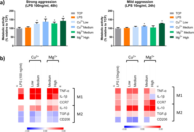
Figure 6.
Effect of the combination of LPS with Cu2+ and Mg2+ on THP-1 cell metabolic activity (a) and gene expression of M1 and M2 markers (b). THP-1 cells were stimulated with a combination of Cu2+ and Mg2+ and 100 ng/ml LPS for 48 h (strong pro-inflammatory stimulation) or 10 ng/ml LPS for 24 h (mild pro-inflammatory stimulation). (a) Metabolic activity results were represented as percentage relative to an unstimulated control (TCP) and compared with the TCP of each day. Statistical significance was accepted at p < 0.05 and represented with *. (b) M1 markers (TNF-α, IL-1β and CCR7) and M2 markers (IL-10, TGF-β and CD206) were determined by RT-qPCR. Results were normalized against β-actin mRNA and are expressed relative to the mRNA levels of unstimulated THP-1 macrophages. Statistical analysis is in Supplementary Tables S4, S5, S6 and S7.

