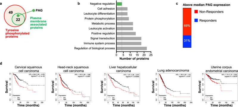
Fig. 1. PAG is associated with negative regulation of biological processes.
a Phosphoproteins were enriched for in the lysates of anti-CD3 or anti-CD3 + PDL2 stimulated Jurkat cells then identified by mass-spec30. 49 proteins were found to be enriched in the anti-CD3 + PDL2 condition relative to unstimulated or anti-CD3. 22 of these proteins were plasma membrane associated, including PAG. b Analysis of the biological processes associated with the 22 plasma membrane associated phosphoproteins. PAG is associated with negative regulation of biological processes (green bar). c Single cell analysis of PAG expression in T cells isolated from cancer patients that received therapeutic PD-1 blockade. Samples are categorized based on patient response to PD-1 blockade (responder eight patients—5110 cells/non-responder 18 patients—10,190 cells) and by median PAG expression level. PAG expression displayed is upper 50%. d TCGA data displayed to show overall survival of high and low PAG expression defined by median expression for each cohort. *p < 0.05.

