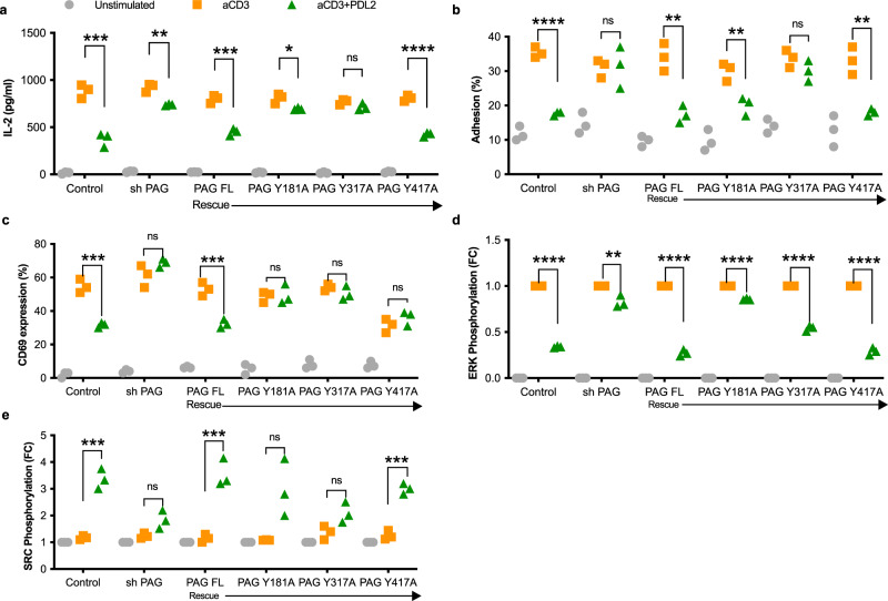
Fig. 3. PAG is required for PD-1 signaling and function.
a ELISA of secreted IL-2 in the supernatants of Jurkat cells collected 24 h following stimulation by magnetic beads. Jurkat T cells either expressed a non-targeting shRNA (control) or PAG targeting shRNA (shPAG). For rescue transfections, shPAG Jurkats were transiently transfected with full length, wild type PAG (PAG FL) or with phosphodeficient mutants (Y181A, Y317A, and Y417A). b Adhesion assay of Jurkat cells to fibronectin following stimulation for 15 min. The number of adherent cells remaining, expressed as a percentage of the total number of labeled cells, was determined with a fluorescent plate reader. PAG knockdown and rescue transfections as in a. c Percentage of Jurkat cells expressing CD69 on the surface following 24-hour stimulation, measured by flow cytometry. PAG knockdown and rescue transfections as in a. d, e Phosphorylated ERK (d) and phosphorylated SRC (e) were detected by western blot of Jurkat lysates 5 min after stimulation by magnetic beads. PAG knockdown and rescue transfections as in a. Fold change is calculated relative to anti-CD3 stimulation (d) or unstimulated (e). Individual data points shown with median indicated of three independent experiments. *p < 0.05; **p < 0.01; ***p < 0.001; ****p < 0.0001; ns not significant.

