Figure 1.
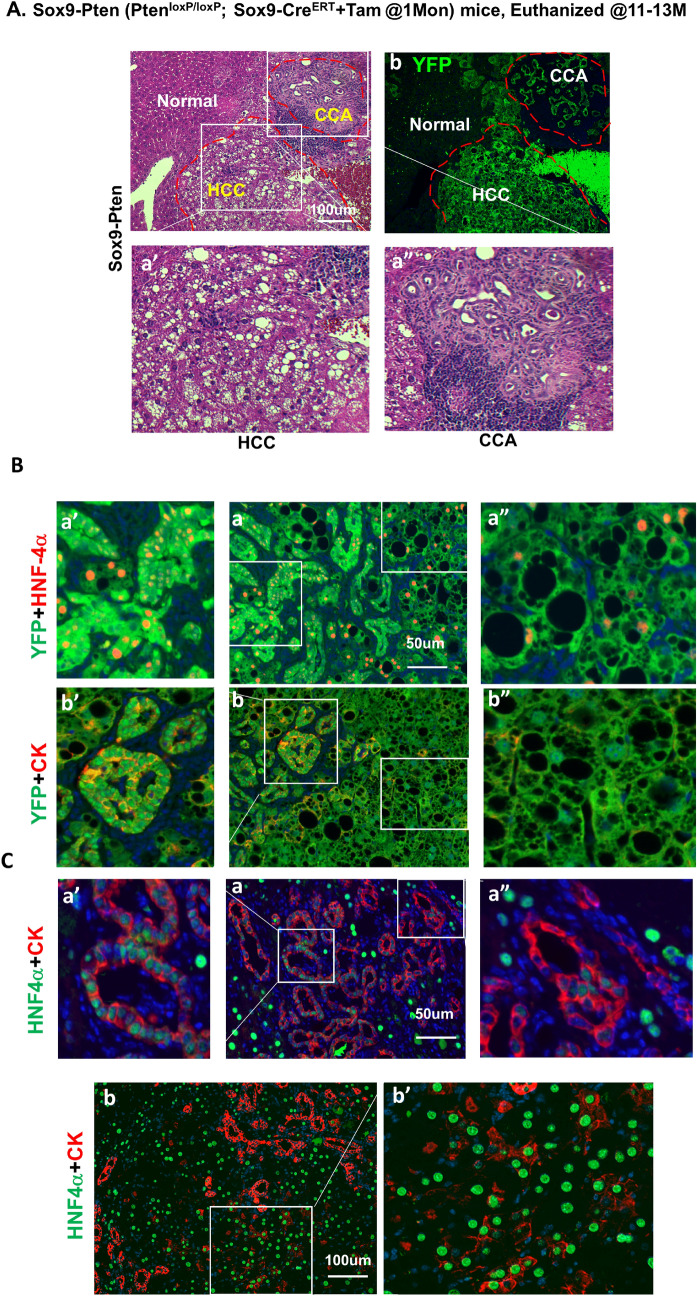
HCC and CCA developed in the aged Sox9-Pten mice. (A) Phenotype of tumors developed in the 11–13 months old Sox9-Pten mice. H&E (a, a′ and a″) and immunofluorescent staining of YFP (b) on serial sections of the tumors developed in Sox9-Pten mice at 11–13 months old. Panels a′ and a″ are amplified views of the boxed areas in panel a. H&E staining shows tumors that morphologically resembles HCC (a′) and CCA (a″). (B) Immunofluorescent staining showing presence of both HCC composed of hepatocytes and CCA composed of cholangiocytes in the YFP positive tumors. Antibodies used are: HNF4α (red) for hepatocytes (a, a′ and a″), CK (red) for cholangiocytes (b, b′ and b″) and YFP. YFP positive cells indicate SOX9+ cells and their progenies that were genetically manipulated to lack PTEN. a′ and b′, enlarged view of morphologically ductal structures within the tumor. a″ and b″, enlarged view of morphologically hepatocyte structures within the tumor. (C) Co-immunofluorescent staining for HNF4α (green) and CK (red) shows tumor cells that express both markers in different areas of the tumor. Panels a, a′ and a″ depict morphological ductal structures that express hepatocytes marker HNF4α (green) in addition to CK (red) which is a marker for cholangiocytes; Panels b and b′ depict morphological hepatic structures that express cholangiocyte marker CK (red) in addition to HNF4α (green) which is a marker for hepatocytes. Blue, DAPI to mark nuclei.

