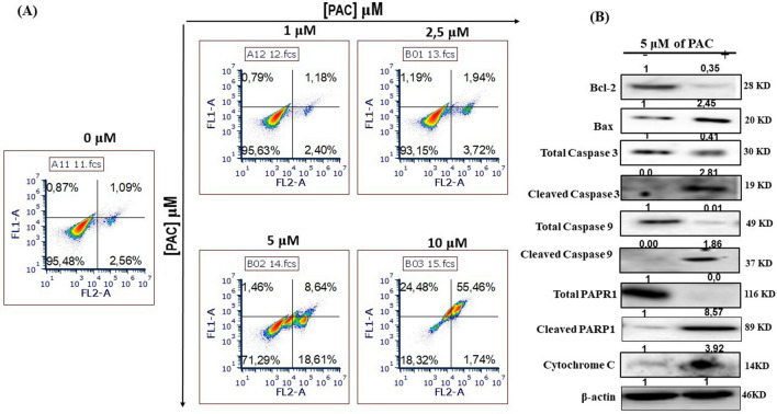
Figure 4.
PAC induces apoptosis in oral cancer cells by targeting the intrinsic mitochondrial pathway. (A) Fluorochrome-labeled annexin V with PI assay; stained cells were subjected to flow cytometry analyses, using a “LSRII” or “CantoII ” cytometer instrument from BD Biosciences (n = 5). (B) PAC dose—dependant increased apoptosis by activation of caspases in Ca9-22 cells (n = 5). (C) The effect of PAC of caspase signaling for oral cancer cell apoptosis was confirmed by Western blotting. PAC at 5 µM strongly decreases Bcl-2 expression and increases that of pro-apoptotic Bax and cytochrome C as well as allows cleavage of PARP-1. The effect of PAC promoting cancer cell death involves caspase-3/9 (n = 3). All Blots for each apoptosis protein derive from the same experiment and were exposed at same time of phosphor imager exposition.

