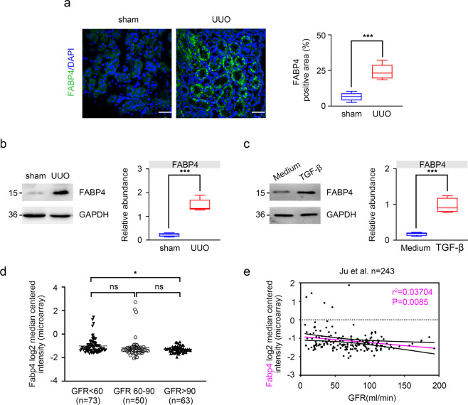
Fig. 1. Expression of FABP4 in UUO mice and TGF-β-treated HK-2 cells.
a Fluorescence microscopy revealed that FABP4 was mainly expressed in kidney tubular epithelial, and increased in UUO-treated mice. Quantitative image to depict fluorescence intensity (Right panel). Scale bars: 50 μm. b Western blot showed that FABP4 was increased in UUO mice. Schematic representation of quantitative data of indicated proteins. Representative images from three independent experiments are shown above. n = 6 mice. c Protein expression of FABP4 was detected in HK-2 cells treated with 10 ng/mL TGF-β for 48 h. Schematic representation of quantitative data of indicated proteins. Representative images from three independent experiments are shown above. d In the study of Ju CKD tubules, CKD was associated with significantly increased mRNA value of Fabp4 compared with biopsy samples from health control. e A negative correlation between tubular FABP4 and eGFR was observed in public Ju CKD dataset. Data were presented as mean ± SEM. *P < 0.05, **P < 0.01, ***P <0.001, ns means no statistical significance.

