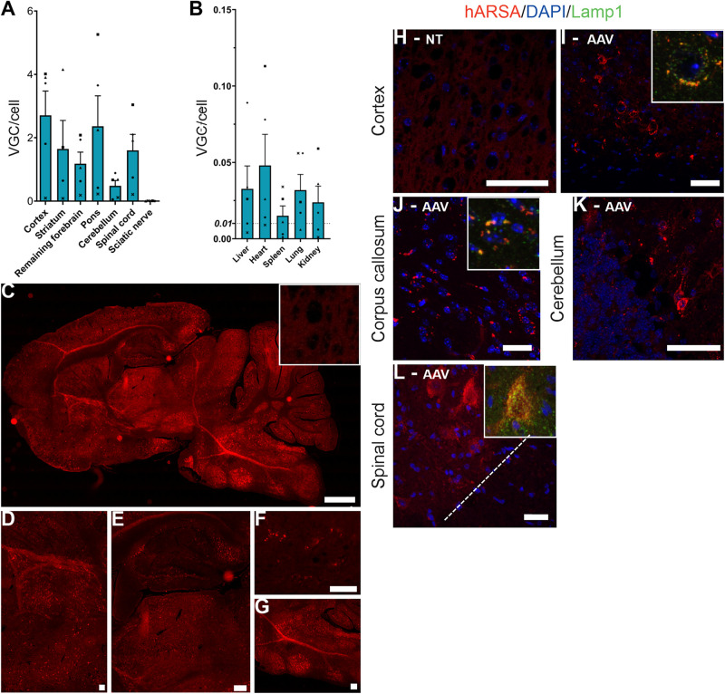
FIGURE 1.
AAVPHP.eB-hARSA-HA efficiently transduce central nervous system. (A,B) Biodistribution of the AAVPHP.eB-hARSA-HA in central nervous system (A) and peripheral organs (B) in 9-month-old KO ARSA mice, 3 months after intravenous injection of AAVPHP.eB-hARSA-HA (n = 5). VGC for vector genome copy number per 2n genome. (C–G) Immunofluorescence detection of hARSA (red) on sagittal sections in the brain (C) and high magnification of different brain areas (D–G) i.e., Striatum (D), hippocampus and thalamus (E), corpus callosum (F) and cerebellum (G) in 9-month-treated KO ARSA mice with AAVPHP.eB-hARSA-HA. Inset is a sagittal section of cortex in control mice. (H–L) Immunofluorescence detection of hARSA (red) and of Lamp1 (green) on sagittal sections in the cortex (H,I), corpus callosum (J) and cerebellum (K) and on coronal sections of spinal cord (L) in 9-month-old untreated (H) or treated (I–L) KO ARSA mice with AAVPHP.eB-hARSA-HA. Nuclei are stained in blue. Insert in (I,J,L) shows a co-localization of hARSA in lysosome (Lamp1, green). Data are represented as mean ± SEM. Scale Bars: 1,000 μm (C); 200 μm (E,G); 100 μm (E) and 50 μm (F,H–L).

