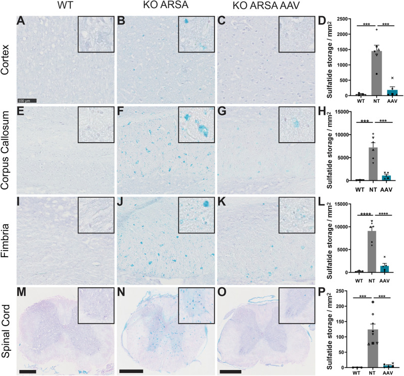
FIGURE 3.
Correction of sulfatide storage in brain and spinal cord of treated KO-ARSA mice, 3 months after treatment (A–C,E–G,I–K,M–O). Alcian blue staining in cortex (A–C), corpus callosum (E–G), fimbria (I–K) and spinal cord (M–O) of wild-type (WT; A,E,I,M), untreated (KO ARSA; B,F,J,N) and AAVPHP.eB-hARSA-HA treated (KO ARSA AAV; C,G,K,O) KO ARSA mice. Inserts are high magnification of tissue section to show the absence or the presence of sulfatide storage. (D,H,L,P) Quantification of sulfatide storage per mm2 in cortex (D), corpus callosum (H), fimbria (L) and spinal cord (P) of WT (n = 3), untreated (NT, n = 6–8) and treated (AAV, n = 5) KO ARSA mice. Data are represented as mean ± SEM. Scale bars: 100 μm expected for (M–O), scale bars: 500 μm. ***p < 0.001; ****p < 0.0001.

