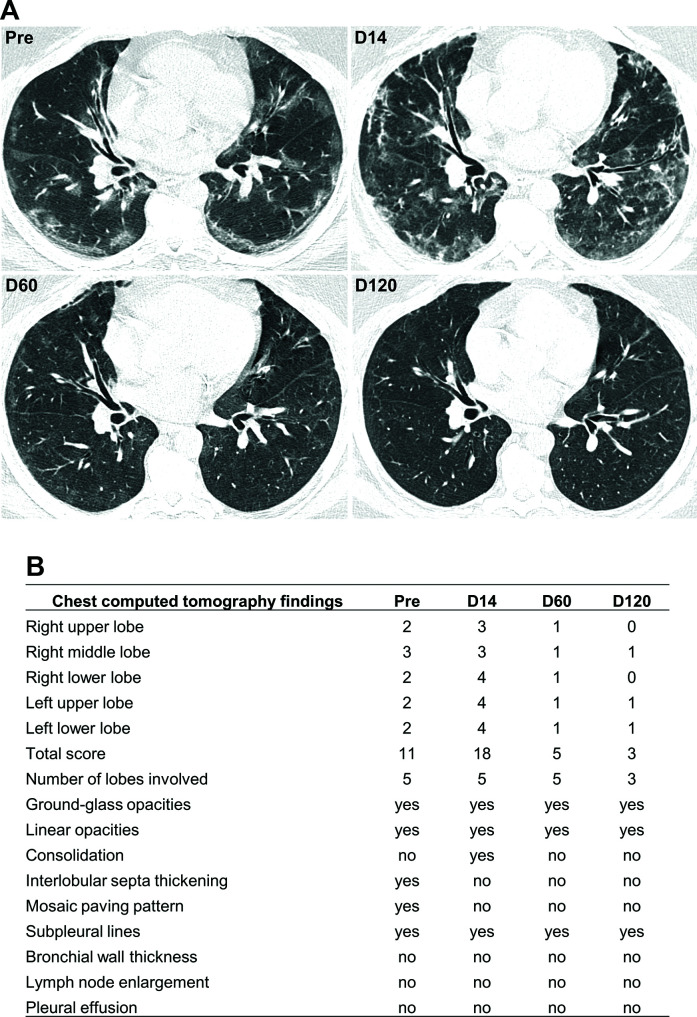
Fig. 5.
Chest computed tomography scan. (A) Chest CT scan without contrast, the axial section at the lower lobes level. Pre-transplantation, showing ground-glass opacities associated with crosslinking (mosaic paving) and subpleural lines, predominantly peripheral and basal. D14 shows an increase in the extent of ground-glass opacities, now with a more significant amount of peribronchovascular opacities. D60 shows marked complete regression of the opacities previously described. D120 shows almost complete regression of the opacities previously described. (B) Each lobe was assigned a score that was based on the following: score 0, 0% involvement; score 1, less than 5% involvement; score 2, 5% to 25% involvement; score 3, 26% to 49% involvement; score 4, 50% to 75% involvement; and score 5, greater than 75% involvement. Scores of 0 to 5 were determined for each lobe, with a total possible score of 25 (adapted from Li et al. 13 ).

