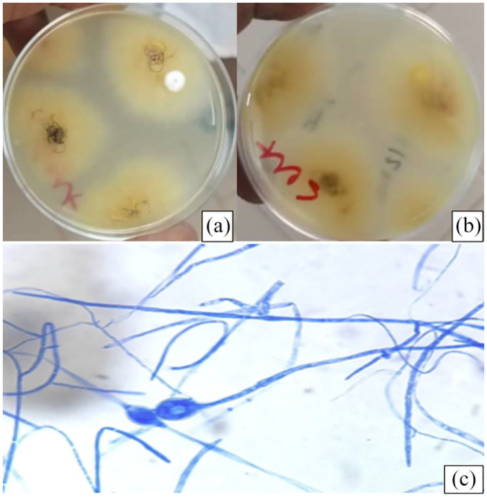
Figure 2.

Colonies of Microsporum audouinii isolated from the scalp lesion. Colonies were yellow-brown to reddish-brown on the front (a) with a beige reverse (b) colonies showing sterile mycelium producing only occasional thick-walled terminal or intercalary chlamydospores (c).
