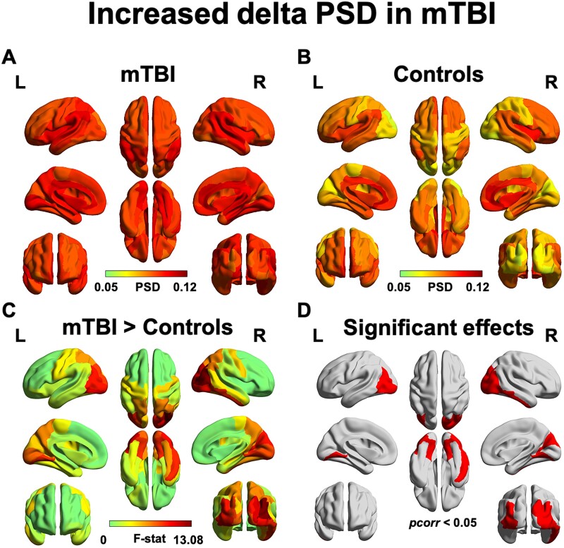
Figure 2.
Children and adolescents with chronic mTBI show pathological increases in delta power. Increased power in the delta band was found in bilateral occipito-parietal areas and the right inferior temporal and somatosensory gyrus. Regional power in the mTBI (A) and the control groups (B) is displayed. The F-statistic map for group contrasts (C) reveal increased power in the mTBI compared to controls as confirmed by a post hoc Wilcoxon Rank-Sum test; binarized FDR-corrected maps showing significant regions (pcorr < 0.05) is plotted in red (D).

