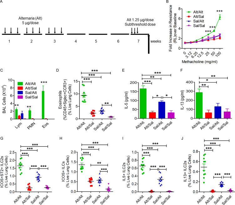
Figure 1.
Establishment of a memory-driven asthma model in Rag1−/− mice. (A) A schematic diagram of the timeline of allergen exposure, recall challenge, and performance of experiments. Groups of Rag1−/− female mice were intranasally exposed to Alt (5 µg in 20 µl of Sal/dose) or Sal three alternate d/wk in weeks 1–3 and then rested for 3 wk. The mice had a recall challenge in week 7 with a subthreshold dose (1.25 µg/dose) of Alt on three consecutive days and then were examined for airway hyperreactivity and immunological alterations 3 d later. (B) Increase in lung resistance over baseline (as measured by flexiVent) in response to increasing doses of inhaled methacholine in Alt/Alt, Alt/Sal, Sal/Alt, and Sal/Sal groups. ***, P < 0.0001 versus control groups, two-way ANOVA, n = 10/group. (C) Differential leukocyte counts of BAL. **, P < 0.001; ***, P < 0.0001, two-way ANOVA, n = 10/group. Eos, eosinophil; Lym, lymphocyte; PMN, polymorphonuclear cell. (D) Eosinophils (CD45+Siglec8+CCR3+ cells) in the lung from the study groups. Blood-depleted lung tissue from the study mice was digested with collagenase, and the single-cell suspension from the lung digest was stained and analyzed for eosinophils by FCM. ***, P < 0.0001, one-way ANOVA, n = 10/group. (E and F) IL5 and IL13 (measured by ELISA) in BAL. ***, P < 0.0001, n = 10/group, one-way ANOVA. (G–J) ICOS+ST2+, ICOS+, IL5+, and IL13+ ILC2s in the lung. Single-cell suspensions from the lung, processed as described above, were stained and analyzed for ICOS+ST2+, ICOS+, IL5+, and IL13+ ILC2s (CD45+CD25+ lung cells) by FCM. ***, P < 0.0001, n = 10/group, one-way ANOVA.

