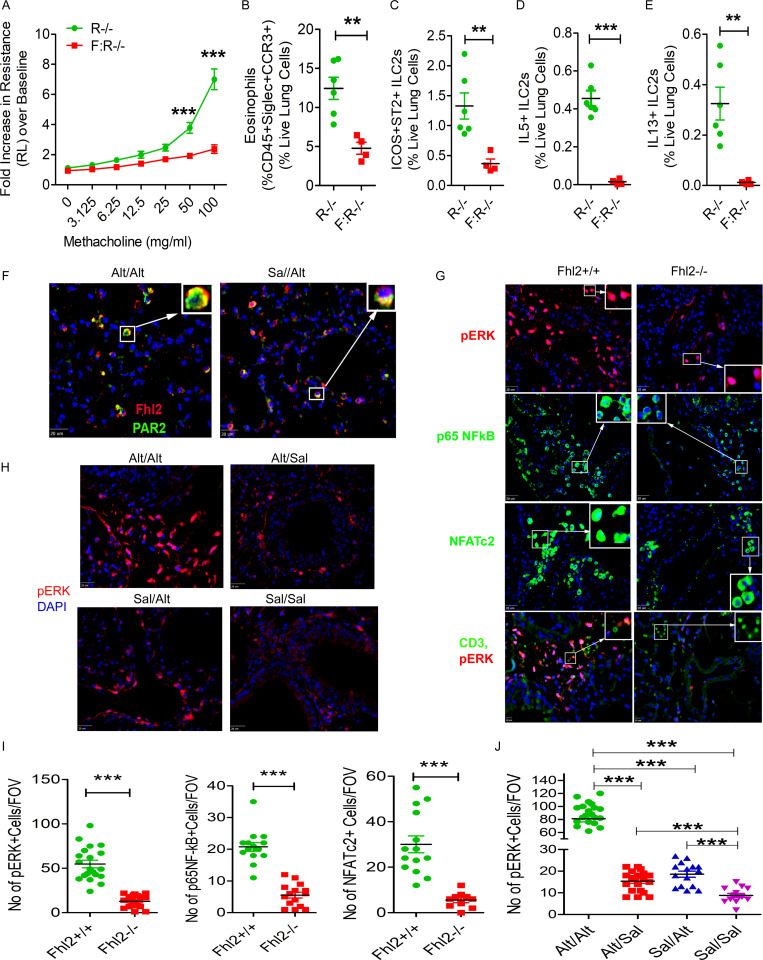
Figure 8.
Importance of Fhl2 in memory-associated signaling. (A–E) Fhl2−/−;Rag1−/− (F:R−/−) mice were sensitized and challenged with Alt and Sal as per Fig. 1 A and then examined for airway hyperreactivity and immunological changes. The increase in lung resistance over baseline (as measured by flexiVent) in response to increasing doses of inhaled methacholine (A), lung eosinophils (B), ICOS+ST2+ ILCs (C), IL5+ ILC2s (D), and IL13+ ILC2s (E). **, P < 0.001; ***, P < 0.001, unpaired t test, n = 5/group. (F) Double immunostaining of the lung tissue from the Alt/Sal and Sal/Sal study group for Fhl2 (red) and PAR2 (green). The insets show a higher magnification of cells displaying colocalization (yellow color; n = 5 mice/group). Scale bars are 20 µm. (G) Immunofluorescence staining of the lung tissue from Fhl2+/+ and Fhl2−/− B6 mice from the memory model for pERK1/2 (red) and p65NFkB and NFATc2 (green). The insets show nuclear localization of NF-κB and NFAT under a higher magnification. Scale bars are 20 µm. The last panel shows costaining of pERK1/2 (red) with CD3 (green). Scale bars are 10 µm. (H) Immunofluorescence staining of the lung tissue from the memory models from Rag1−/− mice for pERK1/2 (red). All sections were counterstained with DAPI (blue), and images were taken at 40×. Scale bars are 20 µm. (I and J) Quantification of pERK+, and nuclear NF-kB+ and NFAT+ cells in the lung tissue from Fhl2+/+ and Fhl2−/− mice (***, P < 0.001, unpaired t test; I) and the quantification for pERK1/2 in memory models from Rag1−/− mice (J). One-way ANOVA. ***, P < 0.0001. FOV, field of view.

