Figure 2.

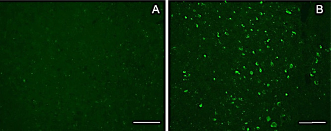
Fluorescence image of brain section from WT (A) and P301S tau mice (B) fixed 1 h after i.v. injection of LM229 showing entry into brain from periphery. Scale bar = 100 μm.

Fluorescence image of brain section from WT (A) and P301S tau mice (B) fixed 1 h after i.v. injection of LM229 showing entry into brain from periphery. Scale bar = 100 μm.