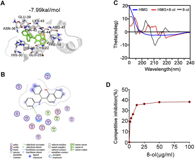
FIGURE 5.
8-ol targets HMGB1 and changes the secondary conformation and disturbs the binding with LPS. (A) 3D binding mode diagrams between HMGB1 and the chemical 7-PMQ-8-ol. The protein is shown in the cartoon and colored in gray, and the chemical is colored in green. The key amino acid residues were shown as sticks. Docking score is −7.99 kcal/mol. (B) 2D binding mode illustrates the key amino acid residues in the ligand–protein complexes. (C) CD spectra of the chemical 8-ol between HMGB1. (D) The LPS-binding capacity of HMGB1 incubated with different concentrations of the chemical 8-ol.

