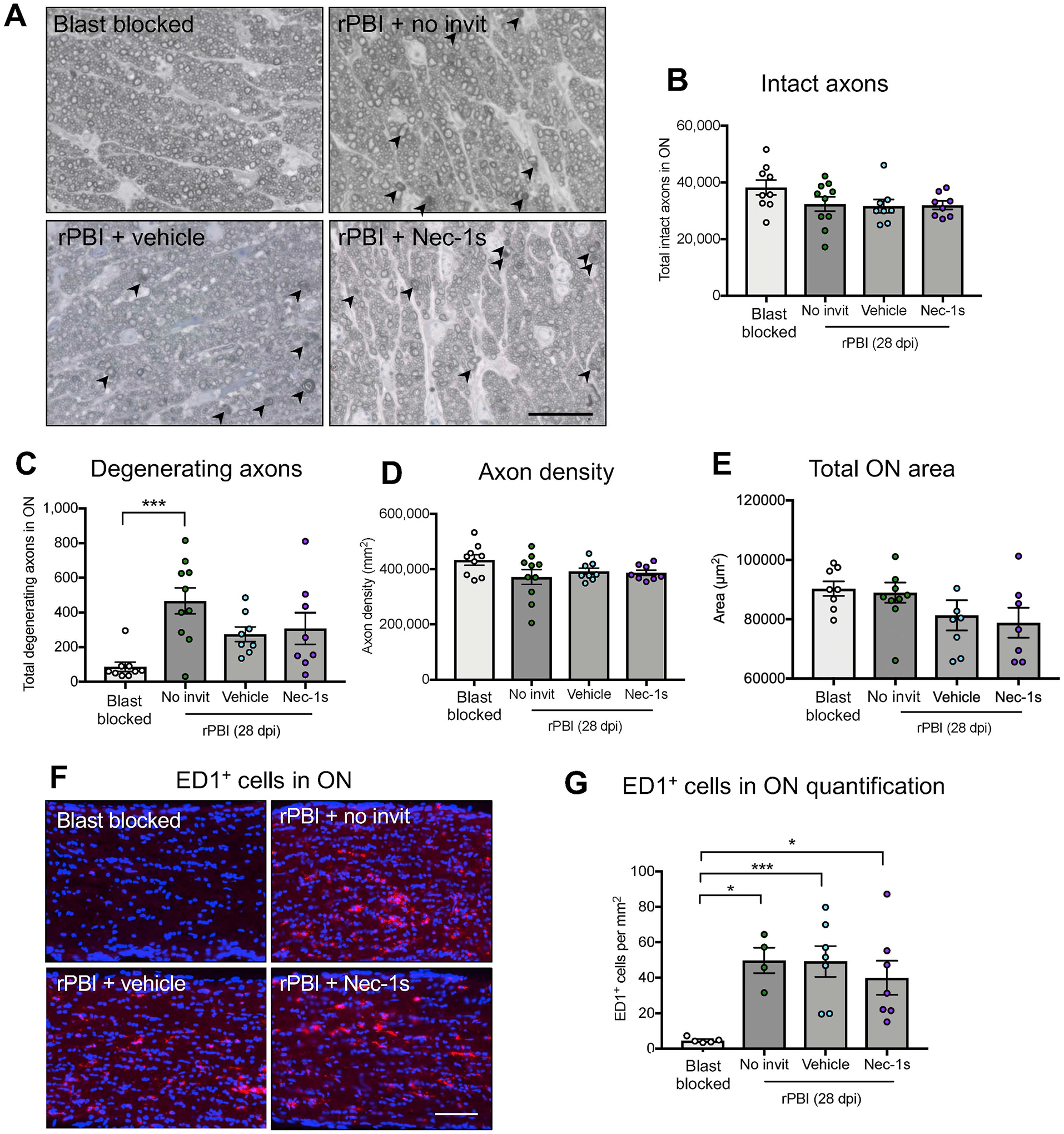
Figure 3. The number of degenerating ON axons and the number of ED1+ cells increased after rPBI.

Total number of intact and degenerating axons were counted in the ON in blast blocked and rPBI treated eyes after intravitreal injections of vehicle and Nec-1s. A) Representative PPD and toluidine blue stained ON semi-thin cross sections showing degenerating axons (arrowheads). B) The number of intact axons was not significantly changed at 28 dpi (P = 0.1729). C) The number of degenerating axons in the ON increased after rPBI compared to blast blocked (P = 0.0014; P = 0.0008 post hoc). There were no differences between Nec-1s or vehicle treated eyes (P = 0.9862, post hoc). D) The axon density was not statistically different 28 dpi (P = 0.4539). E) The total ON area was not changed after injury or Nec-1s treatment (P = 0.3120). Error bars represent mean ± SEM. n = 10 per group. F) Immunostaining of ED1+ cells in the ON. G) The number of ED1+ cells in the ON increased after rPBI with no intravitreal injections compared to blast blocked (P = 0.0057; P = 0.0166, post-hoc) and was also more frequent compared to blast blocked after rPBI with vehicle treatment (P = 0.0064) and Nec-1s treatment (P = 0.0344). Fig. 4A scale bar represents 20 μm and Fig. 4F scale bar represents 100 μm. Error bars represent mean ± SEM. ***P < 0.001, *P < 0.05.
