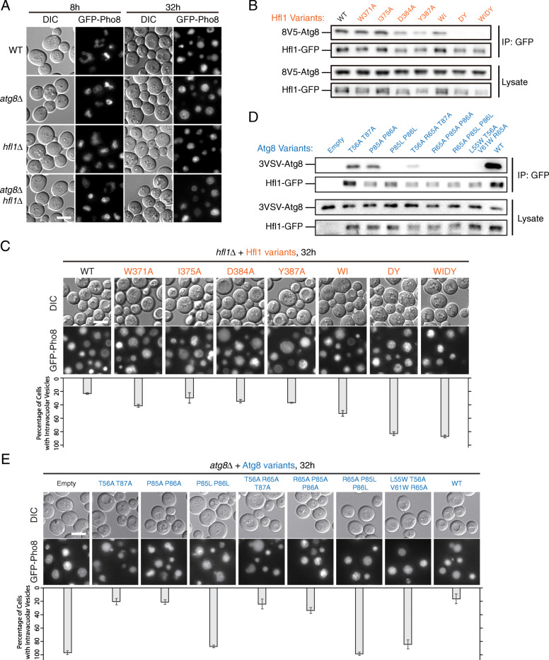
Fig. 6.
Hfl1 and Atg8 function at the same step in EVT. a hfl1Δ cells and atg8Δ hfl1Δ cells accumulate intravacuolar structures similar to atg8Δ cells. Cells treated and image presented as in Fig. 1a. b, c hfl1 mutants defective in Hfl1-Atg8 interaction accumulate intravacuolar structures. WI, W371A I375A; DY, D384A Y387A; WIDY, W371A I375A D384A Y387A. b Assessment of Hfl1-Atg8 interaction by co-immunoprecipitation. In these cells, Hfl1-GFP was expressed under the TEF1 promoter, and 8 V5-Atg8 under its own promoter. Cells in log phase were collected. c Observation of GFP-Pho8 translocation by fluorescent microscopy. Cells treated as in Fig. 1a. Representative images are presented on top. For each strain, at least 160 cells from three independent repeats were analyzed for the presence of intravacuolar structures. The results are presented below the microscopy images. Error bar, standard deviation, n=3. d, e atg8 mutants defective in Hfl1-Atg8 interaction accumulate intravacuolar structures. d Identification of atg8 mutants deficient in Hfl1 interaction. Hfl1-Atg8 interaction was assessed by co-immunoprecipitation as in b. e Observation of GFP-Pho8 translocation by fluorescent microscopy. Cells treated and data presented as in c

