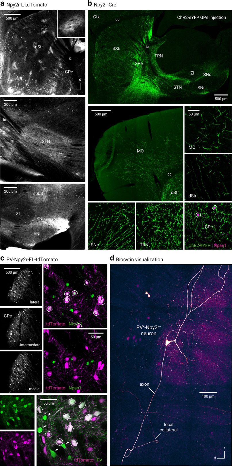
Figure 2.
Npy2r-Cre mice capture a mixed population of GPe neurons. a, Npy2r-L-tdTomato mice labeled neurons in the GPe and striosomes in the dStr (top). Inset, Magnified view of a striosome. Prominent axons were apparent in the STN (middle) and SNc and SNr (bottom). b, Axonal projection patterns from Npy2r+ neurons are similar to that of Npas1+-Nkx2.1+ neurons and Npr3+ neurons. Only a low density of axons was observed in the STN and SNr. Bottom right, ChR2-eYFP+ neurons were largely Npas1+. c, Top left, tdTomato-expressing (PV+-Npy2r+) neurons were observed throughout the GPe in PV-Npy2r-FL-tdTomato mice. PV+-Npy2r+ neurons were immunoreactive for Nkx2.1 (top right) but not for Npas1 (middle right). Bottom, Most of PV+-Npy2r+ (tdTomato+) neurons were PV+. PV+-Npy2r– (green) neurons were visible in the same field (arrowheads). White circles represent colocalization. d, Composite confocal micrograph showing a typical biocytin-filled PV+-Npy2r+ neuron. In addition to the main axon, local collaterals were observed. cc, Corpus callosum; MO, motor cortex; SubB, subbrachial nucleus; ZI, zona incerta.

