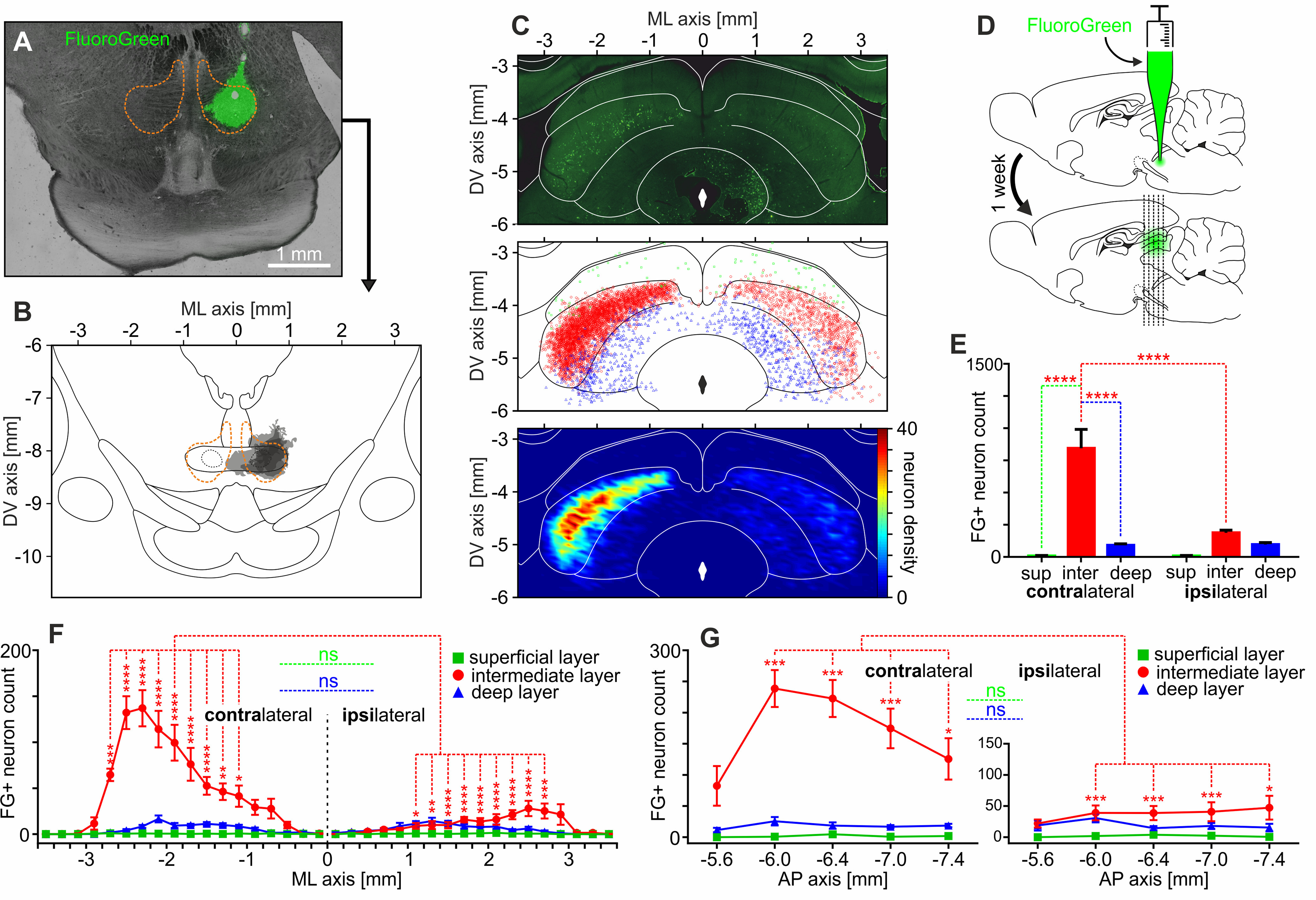
Figure 2.

RMTg is innervated predominantly by the lateral parts of the intermediate layer of the contralateral SC. A, Exemplary image of FluoroGreen injection site within the RMTg. The orange dashed line indicates the RMTg boundaries based on the anti-FoxP1 immunostaining. B, Reconstruction of the FluoroGreen spread at the sites of injections performed in all rats (n = 5). Darker color indicates the overlap of injection across rats. The orange dashed line indicates the RMTg boundaries based on the anti-FoxP1 immunostaining. C, Top, Exemplary image showing retrogradely filled SC neurons after the unilateral injection of FluoroGreen into the RMTg. Middle, Reconstruction of position of retrogradely filled SC neurons observed in all rats (n = 5). Green squares, superficial layers; red circles, intermediate layers; blue triangles, deep layers. Bottom, Density plot showing all retrogradely filled SC neurons. D, Scheme of the experiment. RMTg was unilaterally injected with FluoroGreen, and after 1 week SC was inspected in search of retrogradely labeled neurons. E, Average count of FluoroGreen-positive neurons in both contralateral and ipsilateral SC with regard to SC layers and laterality. F, Distribution of FluoroGreen-positive neurons in mediolateral axis with regard to both SC layers and laterality. G, Distribution of FluoroGreen-positive neurons in anteroposterior axis with regard to both SC layers and laterality. *p < 0.05, **p < 0.01, ***p < 0.001, ****p < 0.0001. ns, Nonsignificant.
