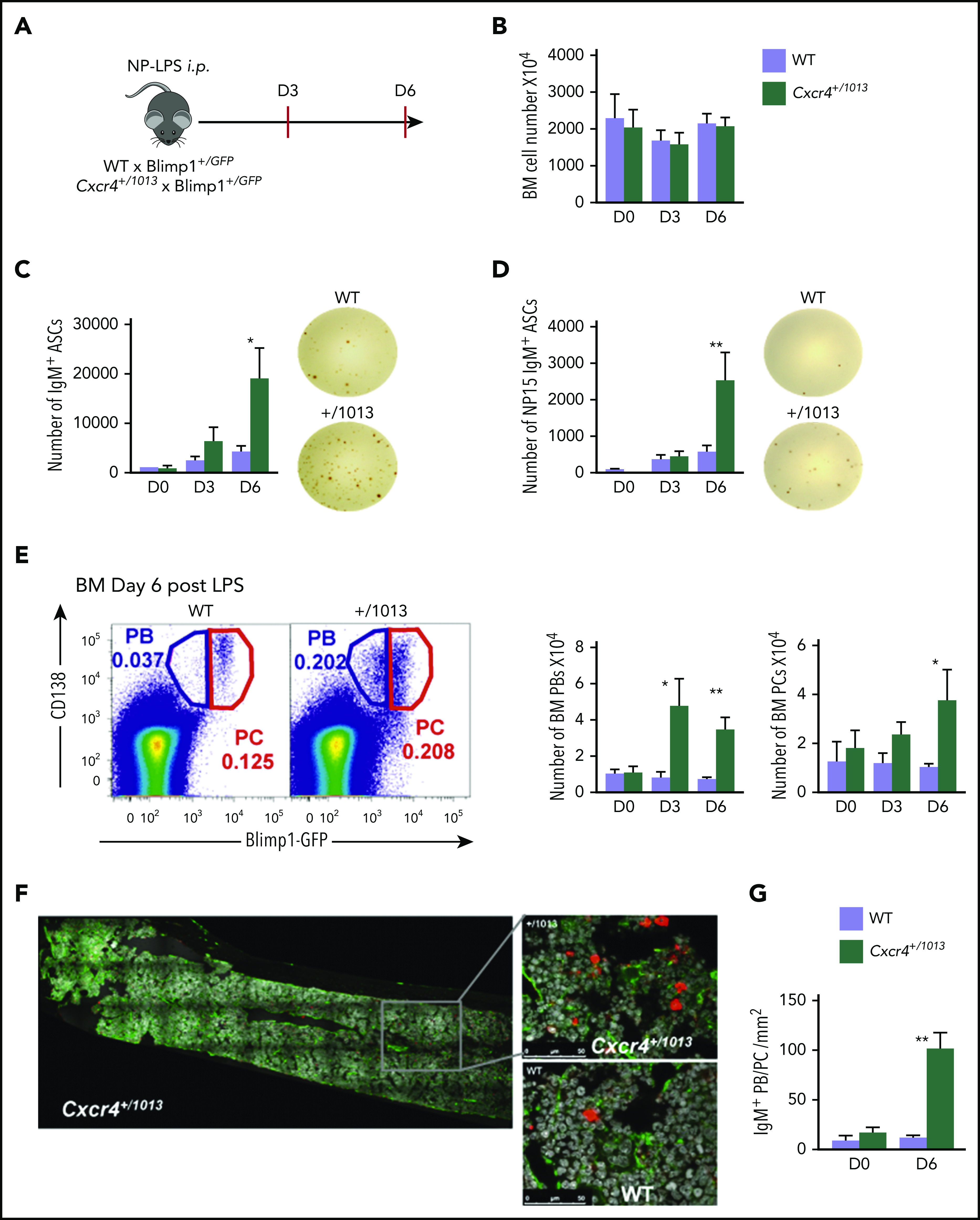
Figure 4.

Fine-tuning of Cxcr4 signaling is required to limit plasmablast accumulation within the BM. (A) Schematic diagram describing the immunization protocol. Blimp1-GFP expression was used to distinguish PBs (Blimp1-GFPlow CD138+) from PCs (Blimp1-GFPhi CD138+). (B) Total BM cellularity of both WT and Cxcr4+/1013 mice during NP-LPS immunization. (C-D) Quantification and representative spots for total IgM+ ASCs (C) and NP-specific IgM+ ASCs (D) in the BM determined by ELISpot at days 0, 3, and 6 after immunization. (E) Representative dot plots and total cell numbers for PCs and PBs in the BM of both WT and Cxcr4+/1013 mice at days 0, 3, and 6 after immunization. (F) Representative images of IgM+ PB/PC staining in BM sections (femur) from WT and Cxcr4+/1013 mice at day 6 after immunization. Whole BM imaging is shown only for Cxcr4+/1013 mice. Sections were stained with Ab against laminin (green) and IgM (red), and nuclei were counterstained with Hoechst 33342 (gray). Scale bars: 50 µm. (G) Quantification of the number of IgM+ PB/PC per mm2 fields at days 0 and 6 after immunization. Results are from 2 to 4 independent experiments (mean ± SEM, n = 5-9 [A-E] or n = 3 [G]). Mann-Whitney U test was used to assess statistical significance (*P < .05, **P < .01).
