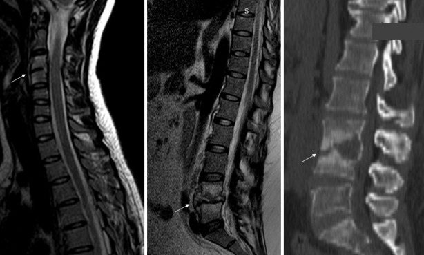
Figure 1.

Example of grade I pyogenic spondylodiscitis (PS) at C3 to C4 level (arrow) on magnetic resonance imaging (MRI; left), grade II PS at L4 to L5 level (arrow) on MRI (center), and grade III PS at L3 to L4 level (arrow) on computed tomography scan (right).
