Figure.
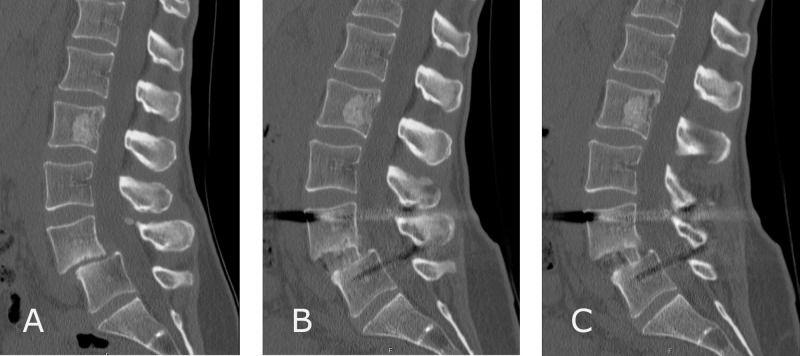
A 47-year-old female patient who presented with spondylolithesis of L4 on L5 in setting of bilateral pars defects (A) and underwent single-level lateral lumbar interbody fusion at L4-5. Six-month postoperative computed tomography scan showed continuous bony union across the interbody cage (B, C).

