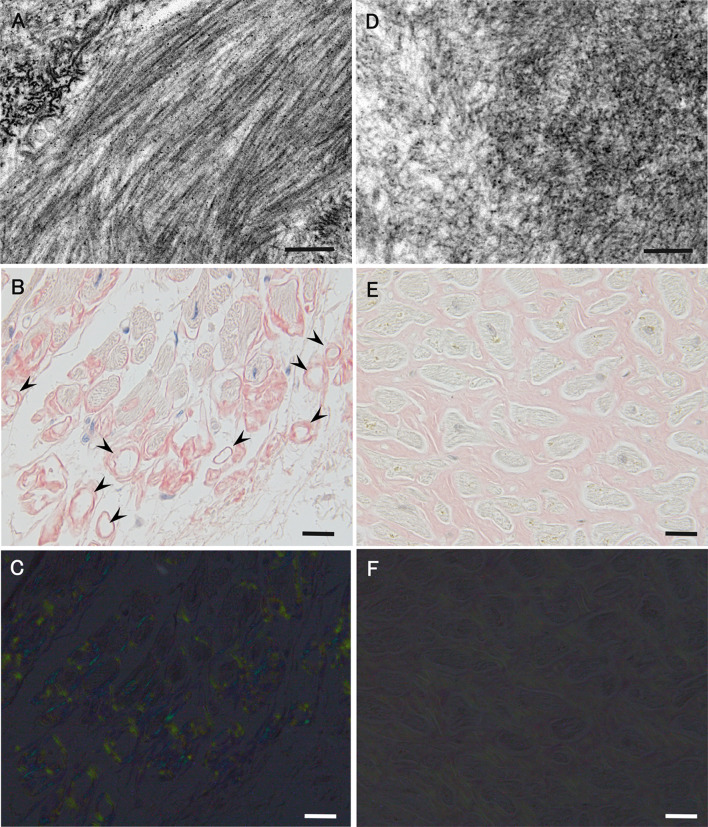
Fig. 7.
Differential characteristics of amyloid deposits between early onset ATTRVal30Met amyloidosis patients from conventional endemic foci (a–c) and late-onset ATTRVal30Met amyloidosis patients from nonendemic areas (d–f). Biopsy specimens of the sural nerve (a, d) and autopsy specimens of the heart (b, c, e, f). Uranyl acetate and lead citrate-stained specimens (a, d). Alkaline Congo red-stained specimens (b, c, e, f). On electron microscopy, amyloid fibrils tend to be long and thick in early onset patients from endemic foci (a). On light microscopy, amyloid deposits tend to be highly congophilic (b) and exhibit a strong apple-green birefringence under polarized light (c) in early onset patients from endemic foci. The atrophy and degeneration of myocardial cells result in the formation of amyloid rings (arrowhead). In late-onset patients from nonendemic areas, amyloid fibrils are generally short and thin on electron microscopy (d). On light microscopy, amyloid deposits are generally weakly congophilic (e) and exhibit a faint apple-green birefringence (f) in late-onset patients from nonendemic areas. Compared with (b), the atrophy of myocardial cells is not conspicuous despite massive amyloid deposition in (e). Scale bars 0.2 μm (a and d) and 20 μm (b, C, e, and f)

