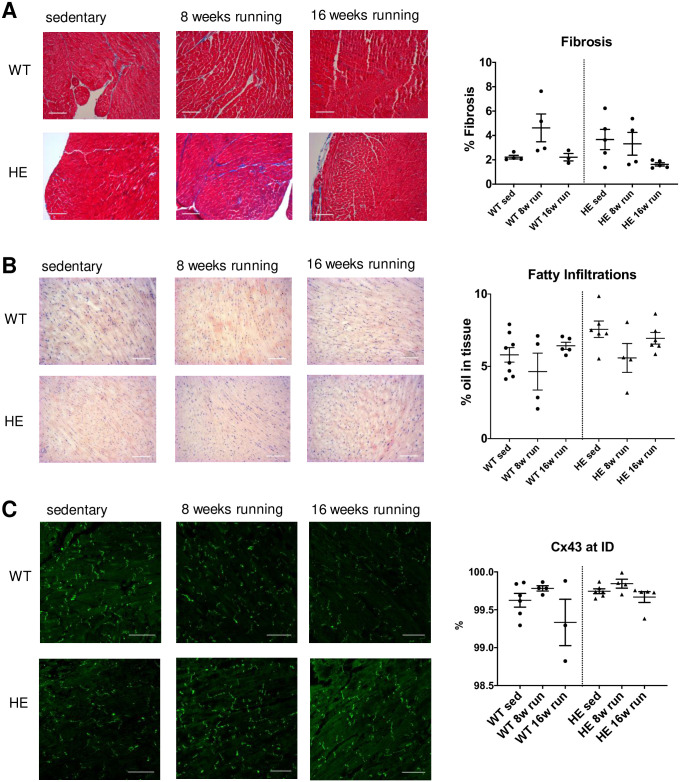
Fig 4. Histological analysis of cardiac tissue after long-term training.
(A) Representative images for sedentary, 8 weeks of running and 16 weeks of running tissue from wt (upper row) and PKP2+/- mice (lower row) are shown for Trichrome-stained ventricular slices. No change of fibrous tissue could be detected in either genotype or in response to long-term training. (B) The oil stain in frozen sections from cardiac tissue with oil red did not reveal an increase in fatty replacements. (C) We labeled the gap junctions with a Cx43 specific antibody and analyzed the images by confocal microscopy. The connexins were predominantly located at the intercalated discs as seen in the representative images shown. There was no shift in subcellular distribution due to genotype or long-term training observed. The single data points indicate the number of animals analyzed.

