
Fig. 1. Genetic and phenotypic AMR.
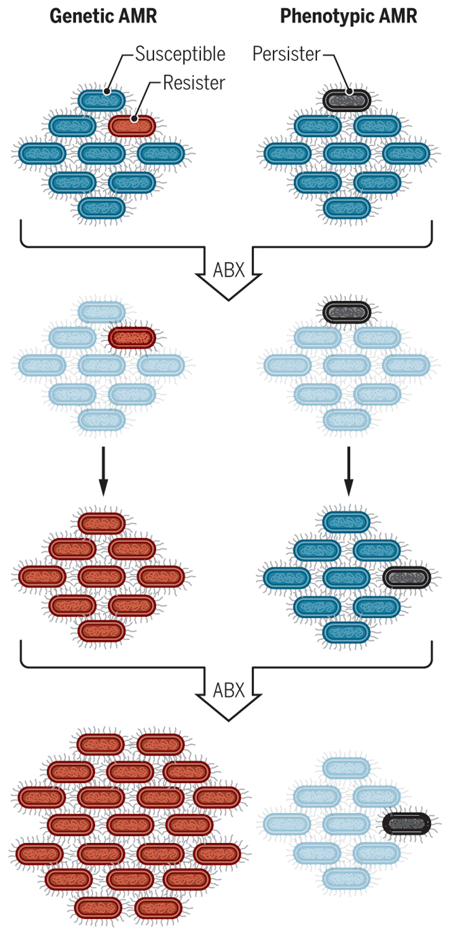
The schematic shows genetic AMR (left) compared to an example of phenotypic AMR (right). Left: In genetic AMR, a bacterium with a mutation that gives it the potential to resist an antibiotic (red cell, first row) survives exposure to that drug (second row) and continues to proliferate, whereas the susceptible majority (blue cells, first row) die (pale blue cells, second row). The resistant bacterium (red) continues to proliferate and pass its mutation on to its progeny even when the antibiotic is removed (third row). Upon a second exposure to antibiotic (fourth row), all the bacterial cells survive and continue to grow during exposure. Right: In contrast, in phenotypic AMR, a bacterial cell is genetically identical to its siblings but happens to be in a metabolic state that is conducive to surviving the first exposure to antibiotic (gray cell, first row). When removed from the antibiotic, this bacterium gives rise to a population resembling that from which it arose so that the second exposure to antibiotic kills the same proportion of the bacterial population as before (fourth row). ABX, antibiotic [adapted from (107)].