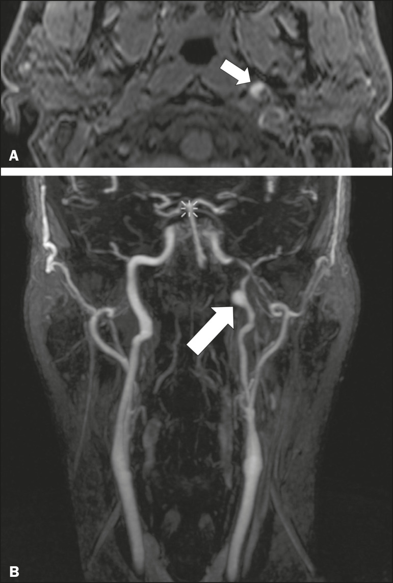
Figure 11.
Carotid artery dissection. A 48-year-old male with sudden left cervical pain accompanied by gustatory changes. Axial T1-weighted fat-saturated MRI sequence (A) and coronal 3D time-of-flight magnetic resonance angiography (B) showing signs of recent dissection of the left internal carotid artery, depicting a crescent-shaped mural hematoma of methemoglobin, causing moderate to severe stenosis of the distal cervical segment. The accompanying focal dilation (0.7 cm) in the upper cervical segment is consistent with a pseudoaneurysm.

