
Fig. 2. In vivo degradation behavior of the PDLLA-Zn-nitrided Fe BRS in rabbit abdominal aortas at different time points after implantation.
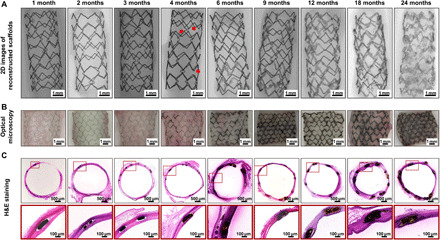
(A) 2D images of micro-CT reconstruction (the red arrows indicate the blurred contour areas at 4 months). (B) Optical micrographs of the scaffolds with vascular tissues. (C) Representative H&E staining images of the scaffolded vessels with high-resolution strut regions.