
Fig. 5. First in-human implantation of the PDLLA-Zn-nitrided Fe BRS.
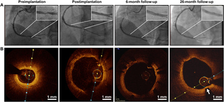
(A) Angiographic appearance of the scaffolded vessel segment and (B) representative OCT images before the procedure, after scaffold implantation, and at 6- and 26-month follow-up.
Official websites use .gov
A
.gov website belongs to an official
government organization in the United States.
Secure .gov websites use HTTPS
A lock (
) or https:// means you've safely
connected to the .gov website. Share sensitive
information only on official, secure websites.
(A) Angiographic appearance of the scaffolded vessel segment and (B) representative OCT images before the procedure, after scaffold implantation, and at 6- and 26-month follow-up.