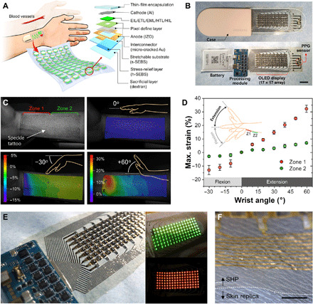
Fig. 1. Concept and structure of a skin-like organic stretchable SHP.

(A) Schematic of an SHP attached to the forearm to measure the heart rate from the wrist (left) and schematic layout of a single pixel on the SRL-embedded stretchable substrate (right). EIL, electron injection layer; ETL, electron transport layer; EML, emitting layer; HTL, hole transport layer; HIL, hole injection layer; IZO, indium zinc oxide; s- and h-SEBS, soft and hard styrene-ethylene-butylene-styrene, respectively. (B) Digital image of a fully integrated standalone SHP comprising an organic stretchable PPG sensor, stretchable OLED display (passive 17 × 7 matrix array), processing module, and thin bendable 3.8-V battery (scale bar, 1 cm). (C) Strain mapping image corresponding to skin deformation and strain distribution according to the wrist angle, as obtained by 3D DIC. A random speckle pattern was formed on the forearm (top left), and the strain distribution was analyzed by monitoring the displacement and deformation of the speckles with a change in the wrist angle between −30° and +60°. (D) Maximum strain experienced by the skin of the forearm at different wrist angles ranging from −30° to +60°. The forearm area near the wrist (12 cm by 4.5 cm) was divided into two equal parts (area, 6 cm by 4.5 cm) designated as zone 1 (part close to the wrist) and zone 2 (part far from the wrist). (E) Digital image of the 15-μm-thick SHP (inset shows green and red OLED arrays). (F) Optical micrographs of the SHP conformably attached to a skin replica (scale bar, 250 μm). Photo credit: Yeongjun Lee, SAIT.
