
Figure 5.
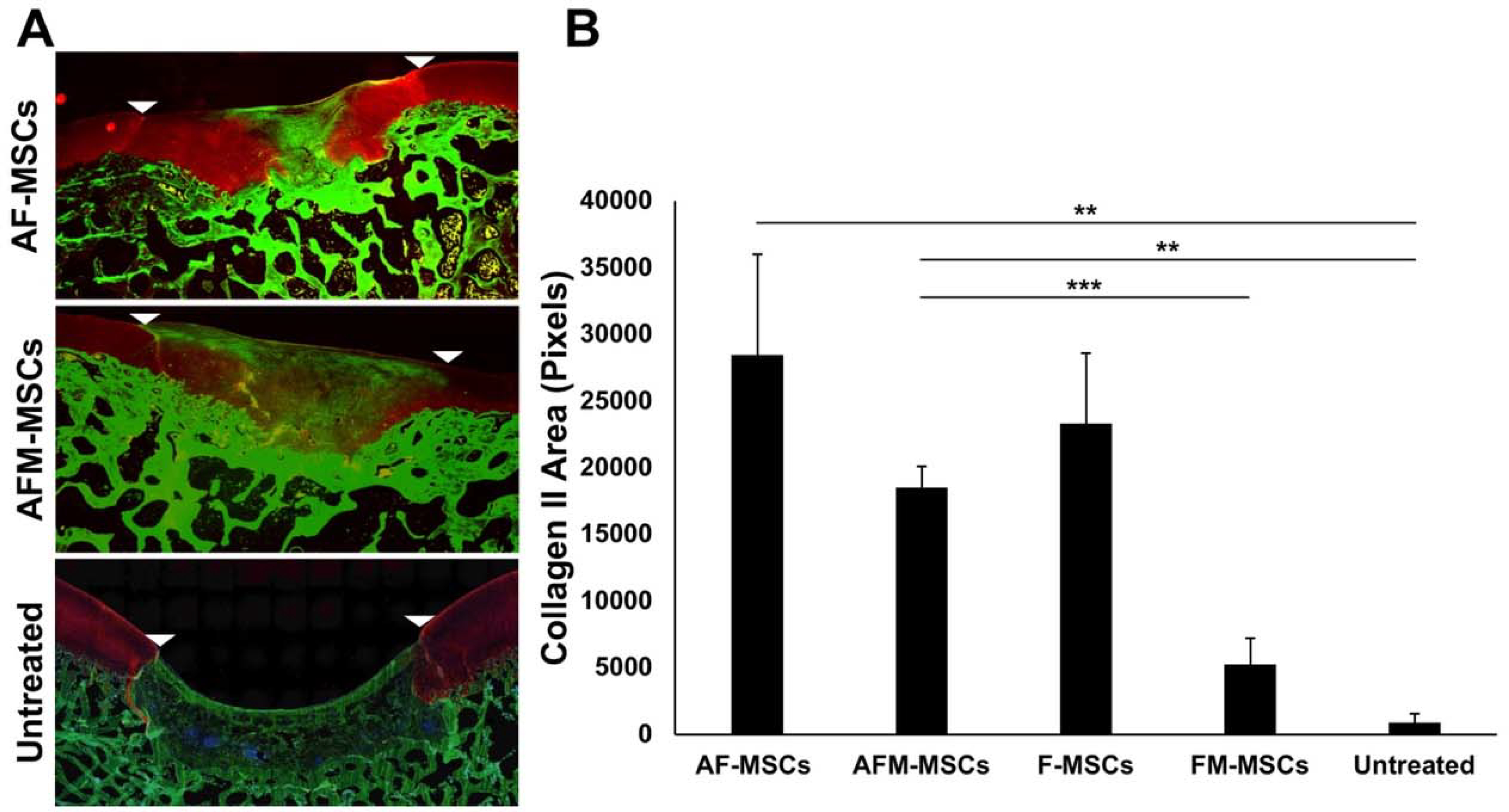
Immunofluorescent staining for collagen II. A) Sagittal sections of the articular cartilage stained with antibodies against collagen II demonstrates marked collagen II production in AF-MSCs and slightly less collagen II in AFM-MSCs. Untreated defects demonstrate lack of collagen II in the defect. White arrow heads indicate defect margins. B) Quantitative analysis of the immunopositive area against collagen II in the repaired cartilage for AF-MSCs (n=8), AFM-MSCs (n=8), F-MSCs (n=10), FM-MSCs (n=10) and Untreated (n=4) group. Data are displayed as means and standard error of the mean. Comparisons between different groups were obtained with Mann-Whitney test. A= Ascorbic acid, F= Ferumoxytol, M= Mitomycin.