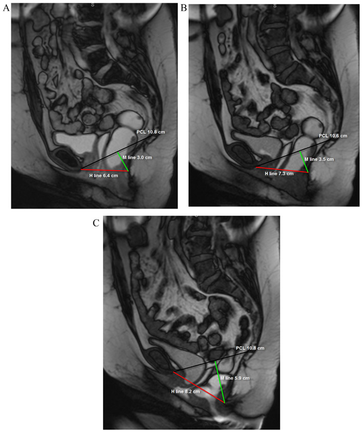
FIG 2.
A 49-year-old female with fecal incontinence. Balanced steady state free precession MR images acquired in static (a) and dynamic phases during Valsalva maneuver (VM) (b) and defecography (c) sequences. The patient demonstrates global pelvic floor instability, with progressive prolapse of the pelvic organs on static, VM and defecography sequences. Note that while the PCL (black line) remains unchanged, there is progressive increase in the H line (red line) and M line (green line).

