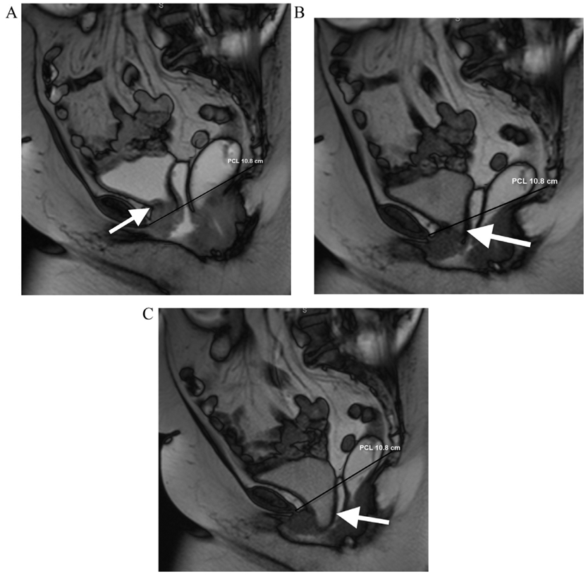
FIG 3.

A 70-year-old female with urinary frequency. Balanced steady state free precession MR images acquired in static (a) and dynamic phases during VM (b) and defecography (c) sequences. The patient demonstrates pelvic floor instability that is worst in the anterior compartment. The bladder base remains above the PCL (black line) on static image (arrow, a), which extending slightly below the PCL on VM (arrow, b). On the defecography sequence, there is marked increase in the degree of bladder herniation below the PCL (arrow, c) demonstrating exacerbation of the cystocele.
