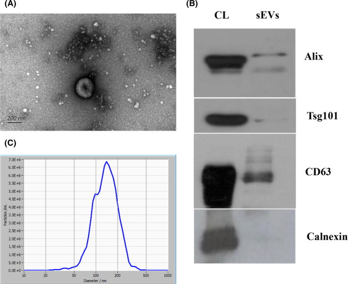
FIGURE 1.

Characteristics of plasma‐derived sEVs. A, Images of transmission electron microscopy showing that sEVs are bowl shaped. Scale, 200 nm. B, Western blot analysis showing the expression of CD63, TSG101, and Alix, and the absence of calnexin. C, Nanoparticle tracking analysis showing the size distribution of sEVs; mean size was 144.6 nm. CL, cell lysates
