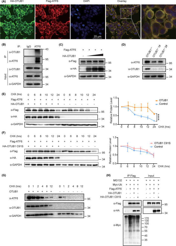
FIGURE 5.

OTUB1 interacts with ATF6 and promotes ATF6 stability. A, Immunofluorescence images of OTUB1 (red) and ATF6 (green) in HEK293T cells. DAPI was used as a nuclear stain (blue). B, Immunoprecipitation experiments showed the endogenous interaction between OTUB1 and ATF6 in T24 cells. C, Cells were transfected with increasing amounts of HA‐OTUB1 (0, 200, 400, or 600 ng) and FLAG‐ATF6, and western blotting was performed to determine the effect of OTUB1 protein levels on ATF6 expression in HEK293T cells. D, Western blot analysis of ATF6 expression in OTUB1‐deficient T24 cells. E, Cells were transfected with FLAG‐ATF6 with or without HA‐OTUB1 as indicated. Western blot analysis of ATF6 stability after treatment with CHX (50 μg/mL) for the indicated time. GAPDH served as a control. The results were plotted (right). F, Cells were transfected with FLAG‐ATF6 with or without HA‐OTUB1 C91S as indicated. Western blot analysis of ATF6 stability after treatment with CHX (50 μg/mL) for the indicated time. GAPDH served as a control. The results were plotted (right). G, Western blot analysis of ATF6 stability in OTUB1‐deficient cells after treatment with CHX (50 μg/mL) for the indicated time. GAPDH served as a control. H, Cells were cotransfected with FLAG‐ATF6 and Myc‐Ub with or without HA‐OTUB1 or HA‐OTUB1 C91S, treated with MG132 (10 μM) for 6 h and then subjected to ubiquitination assays. Data are representative of 3 independent experiments. Statistical significance was analyzed by ANOVA or Student t test. ***<.0001
