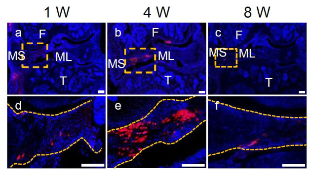
Author response image 3. Representative confocal images of WT mouse knee joints at 1, 4, and 8 weeks after meniscus injury and injection of Gli1+ cells derived from Gli1ER/Td meniscus.
Boxed areas in the top panel are shown at high magnification at the bottom. Dashed line outlines meniscus. Scale bars, 200 μm. Blue: DAPI; Red: Td.

