
Figure 1.
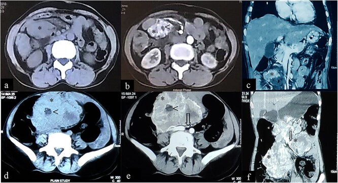
Case 1: CT imaging of extragastrointestinal stromal tumor. (a) Non-contrast axial, (b, c) contrast enhanced axial and coronal reformatted CT images of abdomen demonstrate heterogeneously enhancing confluent mass (*) in peritoneal cavity anterior to the iliac vessels with central area of necrosis (<) peripherally displacing mesenteric vessels (arrow) and bowel. (d–f) Non-contrast axial, contrast enhanced axial and coronal reformatted CT images of abdomen of same patient after 1-year follow-up showing increased interval size of the lesion (*).