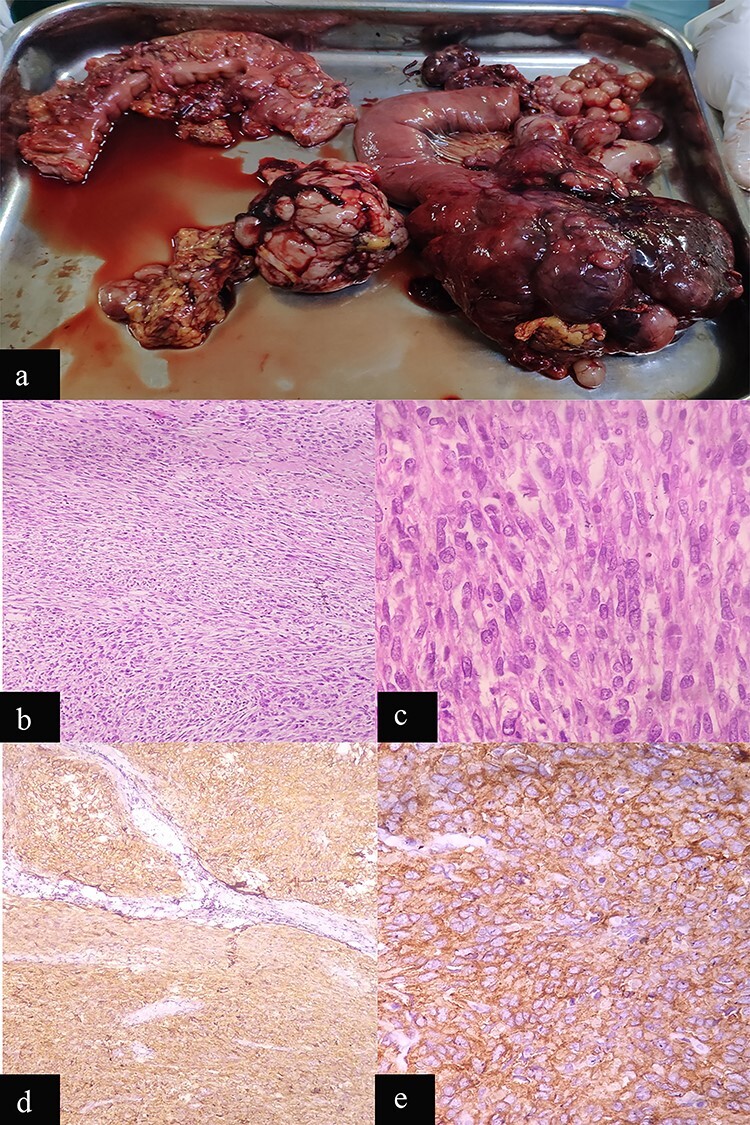
Figure 2.

Case 1: macroscopic and microscopic features of extragastrointestinal stromal tumor. (a) Macroscopic appearance of tumor. (b) Low-power view. Tumor cells are arranged in fascicles. H & E stain. Original magnification ×100. (c) High-power view. The tumor cells have scant to moderate amount of cytoplasm. Nuclei are oval to elongated and have coarse nuclear chromatin. Mitosis is also noted (arrow). H & E stain. Original magnification ×400. (d) Low-power view. IHC showing diffuse staining by CD 117. Original magnification ×100. (e) High-power view. IHC showing diffuse cytoplasmic staining by CD 117. Original magnification ×400.
