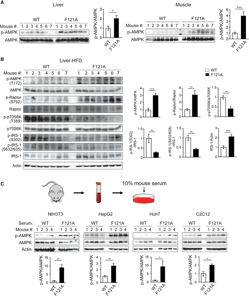
Figure 1. Autophagy-hyperactive Becn1F121A mice show enhanced AMPK activation in tissues, caused by factors in circulation.
(A) Metabolic tissues of Becn1F121A KI mice show increased AMPK activation. Western blot (WB) analysis and quantification of AMPK phosphorylation in liver and muscle of Becn1F121A mice. n = 7.
(B) WB analysis and quantification of AMPK activation and signaling pathways downstream of AMPK in the liver of HFD-fed WT and Becn1F121A mice. n = 7.
(C) Circulating factors in Becn1F121A mice play a role in activating AMPK. WB analysis of AMPK phosphorylation in cell lines cultured for 1 h in medium containing 10% mouse serum from WT or Becn1F121A mice. n = 4.
Data represent mean ± SEM, t test. *p < 0.05; **p < 0.01; ***p < 0.001.

