Figure 1.

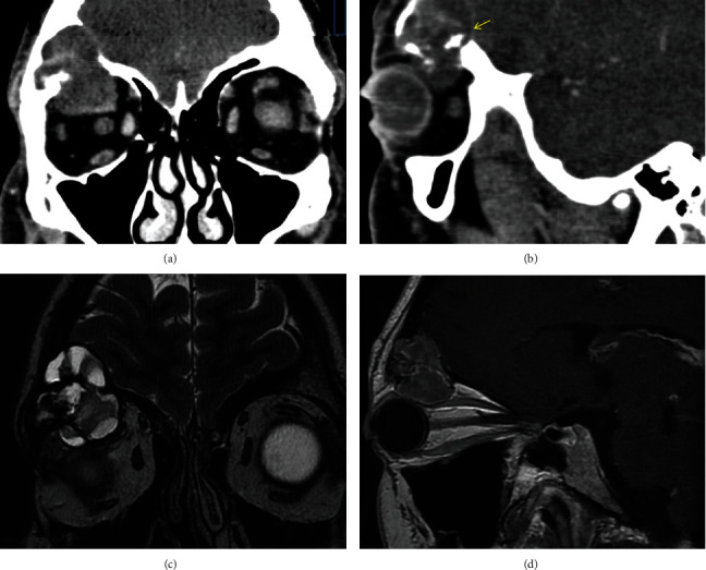
(a, b) Contrast-enhanced coronal and sagittal CT images, demonstrating an expansive lesion arising from the roof of the right orbit with a heterogeneous enhancement and soft tissue attenuation. Inside view of the lesion reveals several small foci of mineralization (arrow). The lesion shows the osseous expansive changes, with thinning of superior and inferior wall of the roof which leads to an invasion over the frontal sinus and into the orbit. (c) Coronal T2-weighted MRI shows a well-defined lesion with a low-signal-intensity margin representing either osseous sclerosis or a pseudocapsule. The lesion shows a multilobulated lytic pattern which reveals markedly increased signal intensity, reflecting the expansive cystic component, and low signal intensity in the small solid regions. (d) Sagittal postcontrast T1-weighted MR image shows a well-encapsulated mass with homogenous contrast enhancement.
