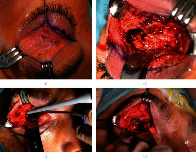
Figure 2.

(a) Surgical approach through an upper eyelid crease incision to access the orbital roof. Intraoperative photograph showing bone destruction in the roof of the orbit and the presence of a red-yellowish mass, with evidence of dark coagulated blood and fragments of bone within the soft tissue. The tumor extends into the anterior cranial fossa. (b) The handpiece of SONOPET® ultrasonic aspirator used for aspiration and emulsification of the tumor tissue in the superior orbit and its extension into cranial cavity. (c) Resection of abnormal tissue from the upper orbit and its extension into anterior fossa.
